larynx
larynx, larynx anatomy, pharynx, larynx anatomy, pharynx anatomy, cuneiform cartilage, larynx, cuneiform cartilage, larynx anatomy, larynx, larynx, larynx anatomy, anatomy of the larynx, pharynx anatomy, ANATOMY OF LARYNX, larynx, cricoid cartilage, laryngeal prominence, larynx, anatomy of larynx,
The following are the result pages for the searched keyowrd: larynx
Larynx Anatomy
19/10/2009 03:06:37 م
In This Section you will find detailed different Photos and images about the anatomy of the Larynx including its surface , attachments , related structures , vocal cords and many more Items about the... More Details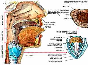
Pharynx and Larynx anatomy
12/10/2009 04:26:00 ص
this is a cut section in the head and the neck showing the laryngopharynx and the larynx on the right is a bigger image of the larynx with its detailed structure showing: 1. post. cricoarytenoid m. 2.... More Details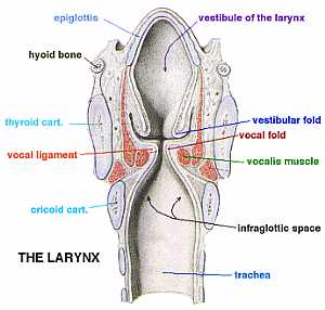
Larynx anatomy
12/10/2009 04:31:00 ص
this image shows the different structures of the larynx showing: 1. epiglottis 2. hyoid bone 3. thyroid cartilage 4. vocal ligament 5. crcoid cartilage 6. vestibule of the larynx 7. vestibular fold 8.... More DetailsLarynx anatomy
12/10/2009 04:31:00 ص
this image shows the different structures of the larynx showing: 1. epiglottis 2. hyoid bone 3. thyroid cartilage 4. vocal ligament 5. crcoid cartilage 6. vestibule of the larynx 7. vestibular fold 8.... More Details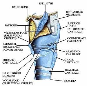
Larynx anatomy
12/10/2009 04:36:00 ص
this is a longitudinal section in the trachea with side view detailing the structure of the larynx showing: 1. epiglottis 2. hyoid bone 3. fat body 4. vestibular fold(false vocal cord) 5. laryngeal pr... More Details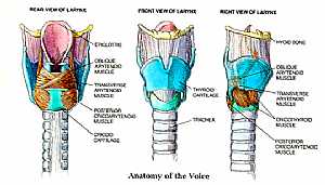
Larynx anatomy
12/10/2009 04:27:00 ص
this is a detailed image of the larynx from ant. and side view showing: 1. epiglottis 2. obique arytenoid m. 3. trans. and oblique arytenoid m. 4. post. cricoarytenoid m. 5. crcoid cartilage 6. thyroi... More DetailsLarynx anatomy
12/10/2009 04:36:00 ص
this is a longitudinal section in the trachea with side view detailing the structure of the larynx showing: 1. epiglottis 2. hyoid bone 3. fat body 4. vestibular fold(false vocal cord) 5. laryngeal pr... More DetailsLarynx anatomy
12/10/2009 04:27:00 ص
this is a detailed image of the larynx from ant. and side view showing: 1. epiglottis 2. obique arytenoid m. 3. trans. and oblique arytenoid m. 4. post. cricoarytenoid m. 5. crcoid cartilage 6. thyroi... More DetailsPharynx and Larynx anatomy
12/10/2009 04:26:00 ص
this is a cut section in the head and the neck showing the laryngopharynx and the larynx on the right is a bigger image of the larynx with its detailed structure showing: 1. post. cricoarytenoid m. 2.... More Details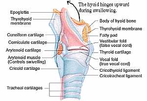
Anatomy of the larynx
11/10/2009 03:44:00 م
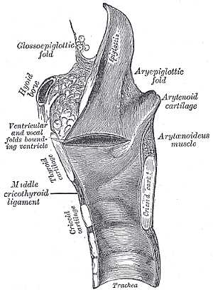
this is the structure of the larynx ( the throat area ) showing: 1. epiglottis 2. thyroid membrane 3. cuneiform cartilage 4. corniculate cartilage 5. arytenoid cartilage 6. arytenoid m. 7. cricoid car... More Details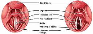
Larynx anatomy
12/10/2009 04:38:00 ص
this is the larynx from above with the vocal cords opened (right image) and closed (left image) showing: 1. base of the tongue 2. epiglottis 3. false vocal cord 4. true vocal cord 5. glottis 6. inner ... More Details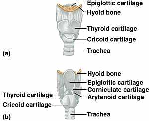
Larynx anatomy
12/10/2009 04:35:00 ص
the upper image is the larynx with thyroid cartilage on the lower image is after removing the anterior part of the thyroid cartilage showing: 1. epiglottic cartilage 2. hyoid bone 3. thyroid cartilage... More Details
Anatomy of the larynx
11/10/2009 03:44:00 م
this is the structure of the larynx ( the throat area ) showing: 1. epiglottis 2. thyroid membrane 3. cuneiform cartilage 4. corniculate cartilage 5. arytenoid cartilage 6. arytenoid m. 7. cricoid car... More DetailsLarynx anatomy
12/10/2009 04:35:00 ص
the upper image is the larynx with thyroid cartilage on the lower image is after removing the anterior part of the thyroid cartilage showing: 1. epiglottic cartilage 2. hyoid bone 3. thyroid cartilage... More DetailsLarynx anatomy
12/10/2009 04:38:00 ص
this is the larynx from above with the vocal cords opened (right image) and closed (left image) showing: 1. base of the tongue 2. epiglottis 3. false vocal cord 4. true vocal cord 5. glottis 6. inner ... More Details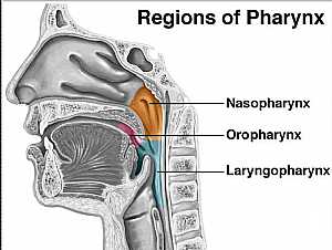
Pharynx anatomy
12/10/2009 04:34:00 ص
this image explains the position of the three parts of the pharynx showing: 1. nasopharynx 2. oropharynx 3. laryngopharynx... More Details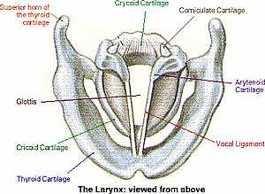
Larynx anatomy
12/10/2009 04:37:00 ص
this is superior view of the larynx showing the vocal cords showing: 1. vocal ligament 2. arytenoid cartilage 3. corniculate cartilage 4. crycoid cartilage 5. sup. horn of thyroid cartilage 6. glottis... More Details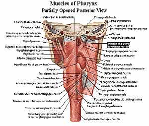
Pharynx anatomy
11/10/2009 04:13:00 م
this is posterior view of the muscles of the pharynx ( which is just the beginning of both the trachea and esophagus) showing: 1. pharyngobasilar fascia 2. accessory muscle bundle 3. digastric m. 4. s... More Details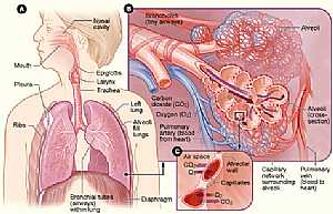
Respiratory system
07/12/2009 07:00:00 ص
this image shows the anatomy of the alveoli and their position in relation to the whole lung and displays their function in the exchange between the blood in the capillaries and the air in the alveoli... More DetailsLarynx anatomy
12/10/2009 04:37:00 ص
this is superior view of the larynx showing the vocal cords showing: 1. vocal ligament 2. arytenoid cartilage 3. corniculate cartilage 4. crycoid cartilage 5. sup. horn of thyroid cartilage 6. glottis... More Detailsorthopaedic joint assessment centr dr mcmahon
, , , , , , ,anatomi ligamen panggul wanita
, ,abdomen sans preparation normale
, , , , , ,world conferences on urine therapy
, , , , , , , , , ,© Copyright 2001-2022 eDoctorOnline.com