trigeminal nerve course
trigeminal nerve, auriculotemporal nerve, trigeminal nerve, trigeminal nerve, olfactory tubercle, maxillary nerve, trigeminal nerve branches, maxillary nerve, trigeminal nerve, trigeminal nerve anatomy, trigeminal lemniscus, olfactory nerve, nasopalatine nerve, trigeminal nerve anatomy, zygomatic nerve, pterygopalatine ganglion, mandibular nerve, mylohyoid nerve, infraorbital nerve, lingual nerve,
The following are the result pages for the searched keyowrd: trigeminal nerve course
Trigeminal nerve anatomy
25/10/2009 03:13:00 م
this is a lateral view of the face with the trigeminal nerve's course indicated on it showing: 1. maxillary nerve 2. mandibular nerve 3. ophthalmic nerve 4. mental nerve 5. labial nerve 6. lacrim... More Details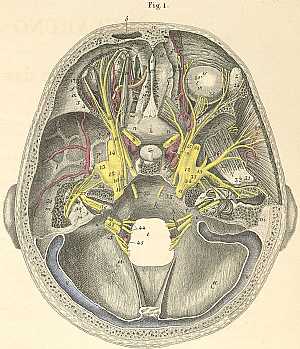
cranial nerves course in the skull
10/10/2009 03:09:00 م
this image shows the course of the cranial nerves inside the skull a) Frontal bone. b) Frontal sinus. c) Internal frontal spine. d) Foramen caecum. e) Crista galli. f) Frontal bone, orbital portion. g... More Details
Cranial nerves anatomy
22/10/2009 01:51:53 م
In This Section you will find detailed different Photos and images about the anatomy of the Cranial Nerves including Their types , Fascial nerve anatomy , trigeminal nerve anatomy , vagus nerve anatom... More Details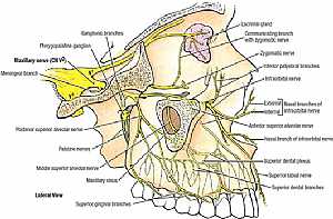
Trigeminal nerve anatomy
28/10/2009 01:55:00 ص
this images illustrates the different branches of the trigeminal nerve in the face in relation to each other [focusing on the maxillary division] showing: 1. maxillary nerve 2. meningeal branch 3. po... More Details
Trigeminal nerve anatomy
24/10/2009 04:17:00 م
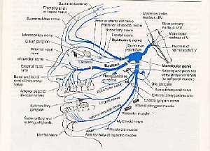
this image shows the course of the different divisions of the trigeminal nerve ( ophthalmic, maxillary and mandibular) showing: 1. ophthalmic nerve 2. mandibular nerve 3. maxillary nerve... More DetailsTrigeminal nerve anatomy
24/10/2009 04:17:00 م
this image shows the course of the different divisions of the trigeminal nerve ( ophthalmic, maxillary and mandibular) showing: 1. ophthalmic nerve 2. mandibular nerve 3. maxillary nerve... More DetailsTrigeminal nerve anatomy
28/10/2009 01:55:00 ص
this images illustrates the different branches of the trigeminal nerve in the face in relation to each other [focusing on the maxillary division] showing: 1. maxillary nerve 2. meningeal branch 3. po... More DetailsTrigeminal nerve anatomy
25/10/2009 03:13:00 م
this is a lateral view of the face with the trigeminal nerve's course indicated on it showing: 1. maxillary nerve 2. mandibular nerve 3. ophthalmic nerve 4. mental nerve 5. labial nerve 6. lacrim... More Details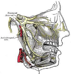
Nasopalatine nerves anatomy
25/10/2009 03:53:00 م
this image shows the course of the nasopalatine nerves (bracnhes of the maxillary division of the trigeminal nerve) showing: 1. trigeminal nerve 2. maxillary nerve 3. ophthalmic nerve 4. mandibular n... More Details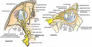
Nerve supply of the eye
28/10/2009 01:50:00 ص
this image shows the nerves supplying the eye in relation to each other from superior view (on the left) and from lateral view (on the right) showing: 1. ophthalmic nerve 2. trigeminal nerve 3. optic... More Details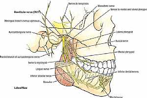
Trigeminal nerve anatomy
28/10/2009 02:07:00 ص
this images illustrates the different branches of the trigeminal nerve in the face in relation to each other [focusing on the mandibular division] showing: 1. mandibular nerve 2. meningeal branch 3. ... More DetailsNasopalatine nerves anatomy
25/10/2009 03:53:00 م
this image shows the course of the nasopalatine nerves (bracnhes of the maxillary division of the trigeminal nerve) showing: 1. trigeminal nerve 2. maxillary nerve 3. ophthalmic nerve 4. mandibular n... More Details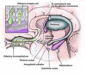
Olfactory nerve course
24/10/2009 04:10:00 م
this image shows the course of the olfactory nerve from the olfactory bulb to centers showing: 1. olfactory neuroepithelium 2. piniform cortex 3. amygdaloid complex 4. entrohinal cortex 5. hippocampu... More Details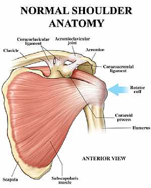
Shoulder joint anatomy
18/12/2009 07:18:00 ص
this image shows the anatomy of the shoulder joint from anterior view displaying the bones, ligaments and muscles in relation to each other. showing: 1. Clavicle bone 2. Coracoclavicular ligament 3. ... More Details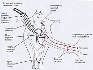
Trigeminal nerve anatomy
28/10/2009 02:44:00 ص
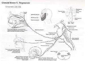
this image shows the origin of the trigeminal nerve at the brain stem showing: 1. mesencephalic trigeminal nucleus 2. spinal trigeminal nucleus 3. pontine trigeminal nucleus 4. motor trigeminal nucle... More Details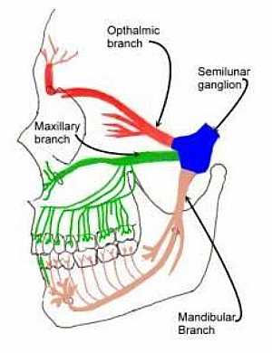
Trigeminal nerve anatomy
28/10/2009 02:41:00 ص
this is a lateral view of the head showing the the different areas supplied by the three divisions of the trigeminal nerve (ophthalmic ,maxillary and mandibular divisions)... More Details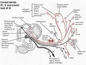
Cranial nerves IX,X,XI anatomy
28/10/2009 02:15:00 ص
this diagram shows the course of the cranial nerves( glossopharygeal IX,vagus X,and accessory XI ) from their origin to the their supplying organs showing: 1. solitary nucleus 2. spinal trigeminal nu... More Details
orthopaedic joint assessment centr dr mcmahon
, , , , , , ,anatomi ligamen panggul wanita
, ,abdomen sans preparation normale
, , , , , ,world conferences on urine therapy
, , , , , , , , , ,© Copyright 2001-2022 eDoctorOnline.com