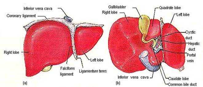
Liver anatomy
this image shows the liver anatomy from anterior and inferior viewanterior view "a" and the inferior view "b"
showing: "anterior"
1. right lobe of the liver
2. coronary ligament
3. inferior vena cava
4. left lobe
5. ligamentum teres
6. falciform ligament
showing: "inferior view"
1. gall bladder
2. right lobe
3. quadrate lobe
4. left lobe
5. cystic duct
6. hepatic duct
7. portal vein
8. caudate lobe
9. common bile duct
10. inferior vena cava
Rate Photo:


16 Ratings
Views: 25825


Link this photo to your website:
Copy the above code and paste it into your webpage, blog or forum
larynx
humerus
corpus callosum
midbrain
neck anatomy
humerus
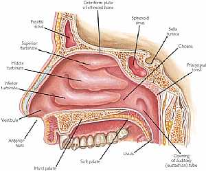
nasal cavity
heart anatomy
midbrain
cranial nerves
eye anatomy
ear anatomy
neck muscles
muscular system
stomach anatomy
stomach
humerus bone
NOSE ANATOMY
Cerebrum
cerebral cortex
FEMUR
anatomy of the neck
humerus bone
eye anatomy
stomach anatomy
pelvic girdle
temporal lobe
Pituitary gland
visual pathway
eye diagram
eye anatomy
femur
falx cerebri
ear anatomy
vagus nerve
neck anatomy
muscular system
heart anatomy
anatomy of neck
eye diagram
lung anatomy
eye anatomy
lung anatomy
nasal cavity
pelvic girdle
visual pathway
brain anatomy
neck anatomy
skeletal system
midbrain
Most Viewed
Most Downloads
 Nose anatomy
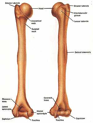 Humerus bone
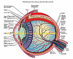 Eye anatomy
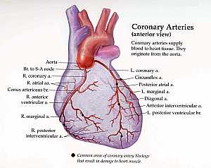 Coronary arteries anatomy
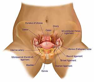 Female pelvic anatomy
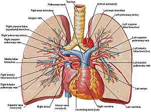 Heart and lung anatomy
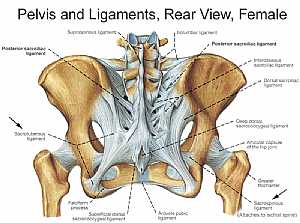 Bones and ligaments of the FEMALE Pelvis
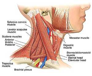 Neck Anatomy
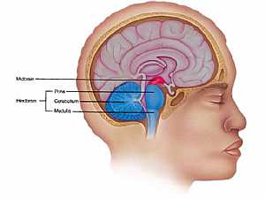 MidBrain anatomy
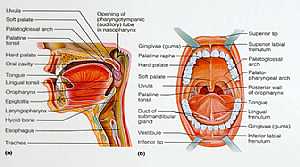 Oral Cavity
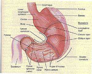 Stomach anatomy
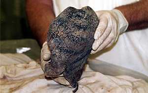 Lung anatomy
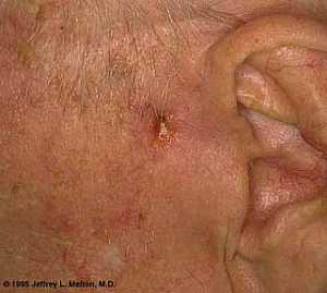 Basal Cell Carcinoma ("Rodent Ulcer" Type)
 Basal Cell Carcinoma ("Rodent Ulcer" Type)
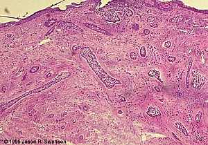 Basal Cell Carcinoma (Histology-Morpheaform Type)
 Basal Cell Carcinoma (Histology-Morpheaform Type)
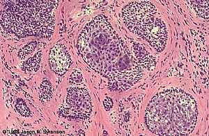 Basal Cell Carcinoma (Histology-Nodular Type - High power)
 Basal Cell Carcinoma (Histology-Nodular Type - High power)
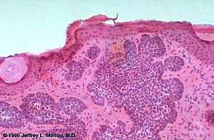 Basal Cell Carcinoma (Histology-Nodular Type- High power)
 Basal Cell Carcinoma (Histology-Nodular Type- High power)
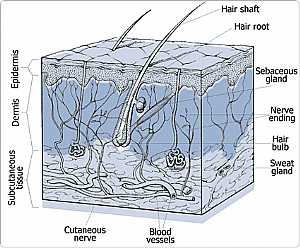 Skin
Skin
 Skin
Skin Nervous System -- Basic
 Nervous System -- Basic
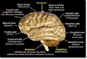 Brain anatomy
 Brain anatomy
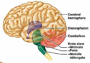 Brain anatomy
 Brain anatomy
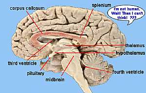 Brain anatomy
 Brain anatomy
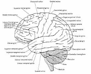 Brain anatomy
 Brain anatomy
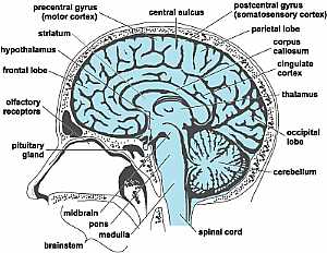 Head anatomy
 Head anatomy
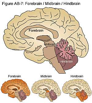 Brain anatomy
 Brain anatomy
eDoctorOnline.com does not provide medical advice, diagnosis or treatment.
© Copyright 2001-2022 eDoctorOnline.com
© Copyright 2001-2022 eDoctorOnline.com
IT IS VREY HELPFULL FOR MY SISTER