Female pelvic anatomy
this image is a horizontal section in a female pelvis shows the different pelvic organs and structures in relation to each othershowing:
1. iliacus muscle
2. sartorius muscle
3. rectus femoris muscle
4. tensor fascia lata muscle
5. vastus lateralis muscle
6. hip joint
7. neck of femur
8. tendon of psoas muscle
9. obturator externus muscle
10. bursa deep to obturator internus muscle
11. ischial tuberosity
12. gluteus maximus muscle
13. obturator internus muscle
14. rectum
15. anococcygeal ligament
16. rectouterine pouch
17. vagina
18. levator ani musle
19. ischiorectal fossa
20. internal pudendal vessels
21. pudendal nerve
22. sciatic nerve
23. posterior femoral cutaneous nerve
24. inferior gluteal artery
25. inferior gluteal artery
26. femoral nerve
27. deep femoral artery
28. femoral artery
29. obturator nerve and vessls
30. femoral vein
31. symphysis pubis
32. bladder
33. pubis
34. superior pubic ligament
35. retropubic space
36. pectineus muscle

Rate Photo:


6 Ratings
Views: 18000


Link this photo to your website:
Copy the above code and paste it into your webpage, blog or forum
larynx
humerus
corpus callosum
midbrain
neck anatomy
humerus
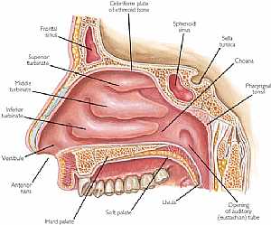
nasal cavity
heart anatomy
midbrain
cranial nerves
eye anatomy
ear anatomy
neck muscles
muscular system
stomach anatomy
stomach
humerus bone
NOSE ANATOMY
Cerebrum
cerebral cortex
FEMUR
anatomy of the neck
humerus bone
eye anatomy
stomach anatomy
pelvic girdle
temporal lobe
Pituitary gland
visual pathway
eye diagram
eye anatomy
femur
falx cerebri
ear anatomy
vagus nerve
neck anatomy
muscular system
heart anatomy
anatomy of neck
eye diagram
lung anatomy
eye anatomy
lung anatomy
nasal cavity
pelvic girdle
visual pathway
brain anatomy
neck anatomy
skeletal system
midbrain
Most Viewed
Most Downloads
 Nose anatomy
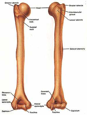 Humerus bone
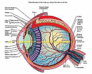 Eye anatomy
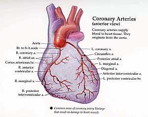 Coronary arteries anatomy
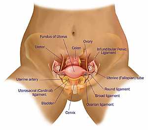 Female pelvic anatomy
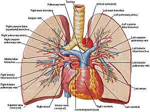 Heart and lung anatomy
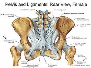 Bones and ligaments of the FEMALE Pelvis
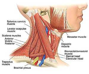 Neck Anatomy
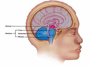 MidBrain anatomy
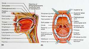 Oral Cavity
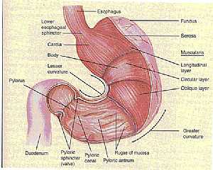 Stomach anatomy
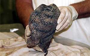 Lung anatomy
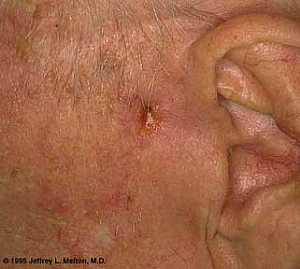 Basal Cell Carcinoma ("Rodent Ulcer" Type)
 Basal Cell Carcinoma ("Rodent Ulcer" Type)
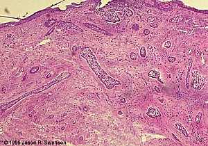 Basal Cell Carcinoma (Histology-Morpheaform Type)
 Basal Cell Carcinoma (Histology-Morpheaform Type)
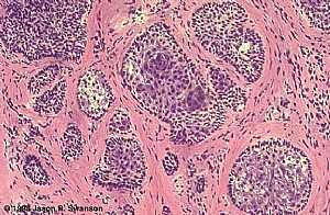 Basal Cell Carcinoma (Histology-Nodular Type - High power)
 Basal Cell Carcinoma (Histology-Nodular Type - High power)
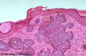 Basal Cell Carcinoma (Histology-Nodular Type- High power)
 Basal Cell Carcinoma (Histology-Nodular Type- High power)
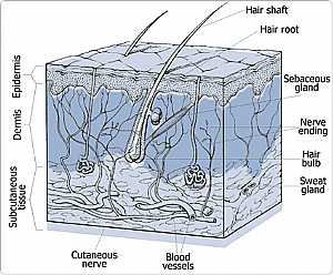 Skin
Skin
 Skin
Skin Nervous System -- Basic
 Nervous System -- Basic
 Brain anatomy
 Brain anatomy
 Brain anatomy
 Brain anatomy
 Brain anatomy
 Brain anatomy
 Brain anatomy
 Brain anatomy
 Head anatomy
 Head anatomy
 Brain anatomy
 Brain anatomy
eDoctorOnline.com does not provide medical advice, diagnosis or treatment.
© Copyright 2001-2022 eDoctorOnline.com
© Copyright 2001-2022 eDoctorOnline.com
I suffer from the disease in the back in the pool
good