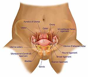
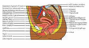
Female pelvic anatomy
this image shows the details of the female pelvis showing the pelvic organs in relation to each othershowing:
1. suspensory ligament of the ovary
2. external iliac artery
3. external ilac vein
4. rectus abdominus muscle
5. subcutaneous fat
6. fallopian tube
7. proper ovarian ligament
8. round ligament
9. uterine lining
10. body of the uterus
11. urinary bladder
12. pubic symphysis
13. urethra
14. clitoris
15. vagina
16. labia minoris
17. labia majoris
18. 5th lumbar vertebra
19. intervertebral disc
20. ureter
21. sacrum
22. spinal cord
23. ovary
24. uterosacral ligament
25. cervix
26. posterior vaginal fornix
27. anterior vaginal fornix
28. rectum
29. urogenital diaphragm
30. anus

Rate Photo:


2 Ratings
Views: 11374


Link this photo to your website:
Copy the above code and paste it into your webpage, blog or forum
Most Popular Tags
labia
,round uterine ligament
, fornix pelvic, fornix vaginae, anterior fornix, minoris anatomy, , ,female photos rectus abdominis and pubic bone
, , , ,female pelvic showing pelvic organ
, , , , , , ,Most Viewed
Most Downloads
 Female pelvic anatomy
 Female pelvic anatomy
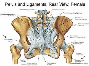 Bones and ligaments of the FEMALE Pelvis
 Bones and ligaments of the FEMALE Pelvis
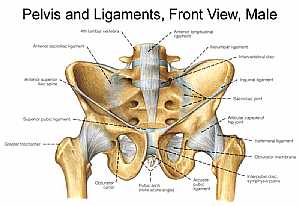 Bones and ligaments of the MALE Pelvis
 Bones and ligaments of the MALE Pelvis
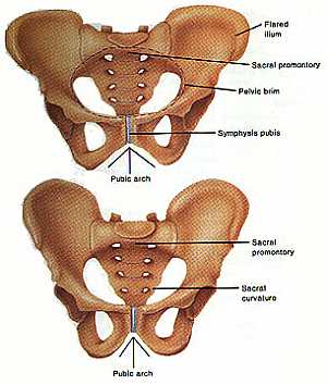 Pelvic girdle anatomy
 Pelvic girdle anatomy
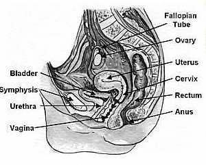 Vagina anatomy
 Vagina anatomy
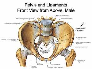 Bones and ligaments of the MALE Pelvis
 Bones and ligaments of the MALE Pelvis
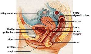 Female pelvic anatomy
 Female pelvic anatomy
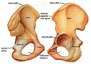 Hib bone anatomy
 Hib bone anatomy
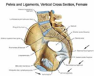 Bones and ligaments of the FEMALE Pelvis
 Bones and ligaments of the FEMALE Pelvis
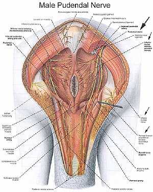 Male pelvic nerves and vessels
 Male pelvic nerves and vessels
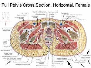 Female pelvic anatomy
 Female pelvic anatomy
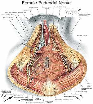 Female pelvic nerves and vessels
 Female pelvic nerves and vessels
 Male pelvis anatomy
 Male pelvis anatomy
Pelvic girdle anatomy
Hib bone anatomy
Bones and ligaments of the FEMALE Pelvis
Bones and ligaments of the FEMALE Pelvis
Bones and ligaments of the MALE Pelvis
Bones and ligaments of the MALE Pelvis
 Pelvic bone and ligaments anatomy
 Pelvic bone and ligaments anatomy
Female pelvic anatomy
 Female pelvic anatomy
 Female pelvic anatomy
 Pelvic nerves and vessels
 Pelvic nerves and vessels
Female pelvic anatomy
eDoctorOnline.com does not provide medical advice, diagnosis or treatment.
© Copyright 2001-2022 eDoctorOnline.com
© Copyright 2001-2022 eDoctorOnline.com
Be the first one to comment on this photo!